In vivo Imaging Facility
- Multi-channel in vivo flow cytometry
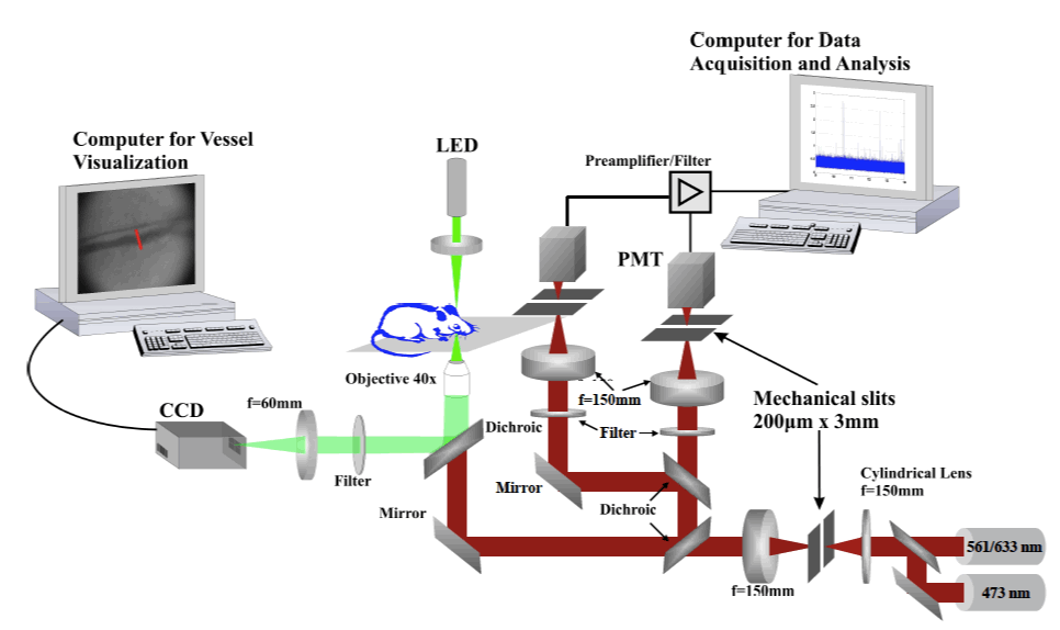
Our group developed a multi-channel in vivo flow cytometer, based on confocal design. Contrary to the conventional flow cytometer, the in vivo flow cytometer can provide real-time detection and quantitative information on fluorescently labeled cells while in circulation in a live animal model. As the labeled cells pass through a slit of light focused across a blood vessel, fluorescence is excited. Its confocal detection makes it possible to observe the cell population of interest without the need to extract a blood sample. Furthermore, the same cell population can be tracked continuously and over long periods of time to examine the dynamic changes in the circulation of different types of cells in the same animal. The in vivo flow cytometer has been used to measure the circulation lifetime of different tumor cells and leukocyte populations in the peripheral circulation in response to immunological stress or therapeutic manipulation.
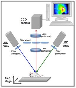
- Whole body imaging system (reflectance mode configuration)
Reflectance imaging is a noninvasive, quantitative method that enables longitudinal studies of physiological changes in individual animals over time. Bioluminescence imaging measures visible light that is emitted by luciferase-driven reactions on the luciferin substrate in the presence of oxygen. Fluorescence imaging quantifies the signal that is emitted by cells and tissues in the animal that have been fluorescently labeled either by transfection (direct insertion of a fluorescent protein promoter into the genome), by direct injection of fluorescence probes or by adoptive transfer of ex vivo isolated fluorescent cell populations. Whole body imaging has been used extensively in cancer imaging, to image the development of implanted tumors in mice, and to monitor metastasis and response to therapeutic intervention. In conjunction with the in vivo flow cytometer, the whole body system has also been employed to investigate the biodistribution and homing kinetics of therapeutic delivery vehicles (microparticles) in animal models. These optical techniques afford the opportunity to quantitatively monitor microparticles in circulation, to detect and image their biodistribution and incorporation in cells and organs of healthy and tumor bearing mice, as well as to acquire anatomical information on cancer size and location.